Homonuclear 2D J-resolved
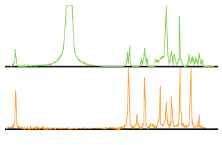
This sequence allows to get the coupling constant along the Fl axis and the chemical shift of each uncoupled proton along the F2 axis (Spectrum 1 and 2). Of course, the couplings involving two different nuclei ( 31P,19F coupled to 1H) are always found in the F2 dimension. One can thus determine the coupling between a proton within a multiplet of a coupled spectrum and phosphorus[1].
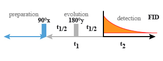
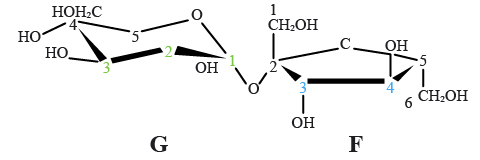
For the 2D J-resolved sequence (Fig. 12), the pulse sequence used is a spin echo sequence. In our example we studied the saccharine molecule (Fig. 29). As in any "echo" experiment, the evolution period
 is divided into two equivalent parts. After the first impulsion of
 , all the magnetization vectors begin to precess as a function of their chemical shifts and the coupling constants.
The impulsion of (180°) on the y axis allows for each refocalised spin to reassociate the vectors with their chemical shifts.
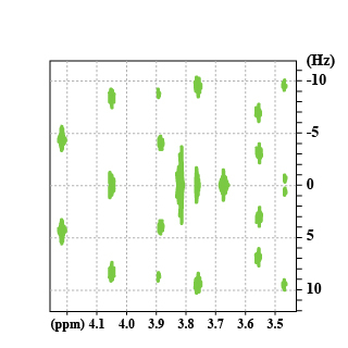
The doublet which is located at 4.22 ppm, shows that the f3 proton has a neighbouring proton. The coupling constant is directly seen on the
 axis. The triplet f4 at 4.05 ppm shows that this one has two protons which are closed to him ... (and so on).